|
|
|
|

|

|
| The Longest Gene | Nov 2002 |
|
What kind of function does the longest gene in the human genome code for? The answer is a
bit mundane: a very long molecular spring that provides muscle with passive elasticity.
Nature adjusts the protein, called titin, for many types of muscle, e.g., skeletal or
cardiac muscle, as well as for other cellular functions. The molecular spring contains
hundreds of elastic elements in series like beads on a string. One type of bead is
the immunoglobulin domain, which can stretch to ten times its normal length. For a long time
only one of the immunoglobulin domains was structurally known, permitting only a single
peek into nature's design library. Recently, a second domain became structurally known
and protein crystallographers and modelers joined forces to discover how nature
designs its beads along titin, as described in a recent publication.
|
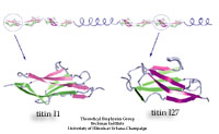
image size:
71.7KB
made with VMD
|
|
Seeking Gold | Oct 2002 |
|
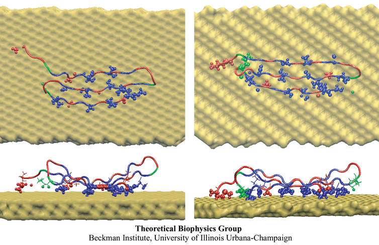
The biological control of inorganic crystal formation, morphology and
assembly is of interest to biologists and biotechnologists studying
hard tissue growth and regeneration, as well as to
materials scientists using biomimetic approaches for control of inorganic
material fabrication and assembly. A molecular-level understanding of
the natural mechanisms involved in these processes can
be derived from the use of metal surfaces to study
surface recognition by proteins together with combinatorial genetics
techniques for selection of suitable peptides.
In a recent study, the structure of a genetically engineered gold binding protein
has been determined computationally, and the ability of the protein to recognize gold crystal surfaces has been explained.
Molecular dynamics simulations were carried out with the
program NAMD using the solvated protein at the gold surface to
assess the dynamics of the binding process and the effects of surface
topography on the specificity of protein binding.
|
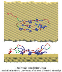
image size:
91.6KB
made with VMD
|
|
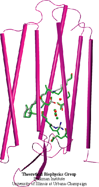
Nailing the Mechanism of a Protein | Sep 2002 |
|
Resolving the physical processes that underly the biological function of a
protein can be an elusive goal even with extremely detailed observations.
An example is the protein bacteriorhodopsin, a light driven proton pump in
archaebacteria. This protein is a close relative to human G-protein
coupled receptors that are the target for many pharmacological
interventions and, hence, knowledge of bacteriorhodopsin's dynamics is of
great medical interest. Despite the availability of highly resolved
structures and spectroscopic observations of the protein and its
functional intermediates, as they arise within 10-12 s of light
absorption triggering its function, the physical mechanism remained ill
understood. A recent computational modeling study
that combined a quantum mechanical simulation of the protein's active site
with a classical mechanical simulation of the remainder of the protein
succeeded to fill in the elusive detail that reveals a complete picture of
how the protein initiates proton pumping, a key step to explain entirely
the biological function. For more information see here.
|
image size:
139.9KB
made with VMD
|
|
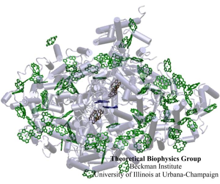
Unbreakable Biological Solar Cell | Aug 2002 |
|
Light is fundamental for life. Through many
photosynthetic life forms, its energy fuels the major part of Earth's
biosphere. The familiar green color of plants, so ubiquitous in our
surroundings, stems from chlorophylls, molecules that help plants, algae,
and some bacteria to harvest the sunlight. Recently, the structure of an
apparatus that harvests sunlight in cyanobacteria, and actually in a
similar fashion in plants, has been discovered, showing 96 chlorophylls
being held at close distances by a protein complex. The chlorophylls
absorb sunlight and deliver its energy to a central chlorophyll pair that
utilizes it to electronically charge a cell membrane, the whole
functioning like an extremely efficient biological solar cell. Quantum
physics and a theoretical analysis of the energy utilization of the
system, reported in a recent
publication, have revealed that this system has been designed with a
high degree of fault tolerance and optimality: pruning single and even
multiple chlorophylls hardly affects the efficiency of the apparatus;
altering the chlorophylls' arrangement though leads to a reduction of
efficiency. Since the apparatus is naturally exposed to intense radiation
and subject to continuous damage, its robustness is crucial for the
organism.
|

image size:
62.3KB
made with VMD
|
|
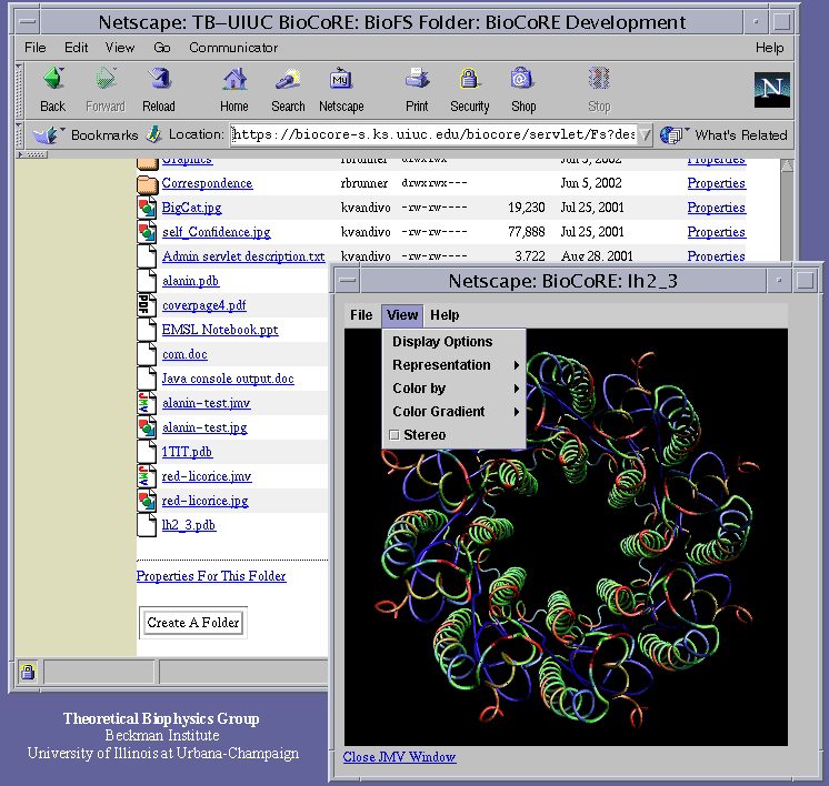
Proteins Through the Looking Glass | Jul 2002 |
|
The building blocks of living cells are biomolecules so small that no light microscope can see them, yet viewing them is essential to decipher the inner workings of cells. The best looking-glass for biomolecules (such as proteins) available today is computers running molecular graphics software that translates experimental data into the molecular graphics. Now the wide availability of molecular graphics has taken a step forward with our new visualization package, JMV (Java Molecular Viewer). JMV borrows several key features from our visualization tool for large scale biomolecules, VMD. The JMV applet places the picture of a protein in your web browser, shown in a 3-D view, ready to be rotated, scaled, and colored according to physical properties. JMV will serve the next generation of bioinformatics web tools, like BioCoRE, through its great adaptability and will turn every molecular picture in electronic text books or web sites into an interactive looking-glass.
|
image size:
379.0KB
|
|
A Molecular Sieve | Jun 2002 |
|
Living cells rely on nutrients absorbed through their cell membranes, for
example on glycerol that is key to the cells' metabolism. Proteins,
so-called aquaporins,
in the membranes form channels that act as sieves
permitting passage of water, glycerol, and like molecules, but prevent
other molecules of similar size from entry and clogging. For this purpose
the channels interact strongly with molecules attempting to pass. In a
recent
publication, the energetics of the conduction process of
glycerol for the aquaporin GlpF was measured in a molecular dynamics
simulation, carried out with NAMD, that pulled
glycerol through the channel, monitoring the forces needed to advance
along the channel axis. An analysis that discounted the irreversible work
done on glycerol, a difficult prerequisite, yielded the energy profile
that glycerol experiences along the channel and that reflects how the
protein decides which molecules are allowed to pass the sieve.
|
image size:
104.1KB
made with VMD
|
|
Filtering a Bathtub of Water a Day | May 2002 |
|
Human kidneys need to filter about a bathtub of water a day through
cells that contain membrane channels made of proteins called aquaporins.
Crystallographers from the University of California at San Francisco (R. Stroud and
coworkers) that discovered the structure of one type of aquaporins,
aquaglyceroporins, have teamed up with UIUC researchers to determine how
these channels achieve their very high water throughput, yet prevent the
cells' electrical potential from discharging by not permitting any flow of
ions or conduction of protons. The team, combining 106,000 atom
simulations using NAMD and crystallography,
found that the channels achieve the impressive filtering function by
conducting water single file and keeping the water molecules strictly
oriented: water molecules enter the channel oxygen atom first and leave the
channel oxygen atom last. Aquaporins are ubiquitous in mammals, plants,
and bacteria and the finding, published recently in Science magazine,
has implications for many biological functions as well as for human
diseases, e.g., cataract of the eye, loss of hearing, or diabetes
insipidus. (more, press
release)
|
image size:
82.5KB
made with VMD
|
|
Forceful Signaling | Apr 2002 |
|
Biological cells process numerous types of information, for
optimal control of their life cycles or to adapt to their environment,
and recruit for this purpose signaling proteins. The best known among
the latter are the G-proteins, involved in numerous diseases and
related to many targets of drugs. G-proteins are closely related to
motor proteins: G-proteins get switched on and off through the binding
of GTP and its hydrolysis to GDP; motor proteins generate mechanical
force through binding of ATP and its hydrolysis to ADP. A recent publication reports a 19,463 atom computer simulation
using NAMD that reveals a "power
stroke" in G-proteins likewise found in motor proteins. The stroke
switches on and off G-proteins' ability to interact with other
signaling proteins, with a power stroke that transforms the protein
from an ordered into a disordered conformation.
|
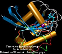
image size:
98.9KB
made with VMD
|
|
Exciting Biology and Hot Physics Meet | Mar 2002 |
|
Most life forms exist near temperatures of about 300 Kelvin where
thermal disorder is significant. Understanding how life copes with
this disorder, in fact, most often exploits it, poses a deep
intellectual challenge. Two recent publications investigate thermal
disorder for electronically excited bioelectronic systems that harvest
sun light and funnel
its energy into the metabolism of so-called purple bacteria. One study
borrows mathematics (supersymmetric calculus) from the physics of
elementary particles to describe the optical properties of randomly
distributed, but otherwise immobile, aggregates of chlorophylls. The second
study
goes a step further and investigates optical properties
affected by thermal motion. The paper draws its insights from a
pioneering 87,055 atom molecular dynamics simulation of a
membrane-protein-chlorophyll system that monitored thermal motion of
atoms and electrons and extends a mathematical description, the
polaron model, used in advanced solid state physics.
|
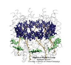
image size:
122.6KB
made with VMD
|
|
The First 0.000000000001 Second of Vision | Mar 2002 |
|
Since Newton, vision has attracted physicists seeking to explain how
light is sensed by organisms.
Recently, the structure of a visual receptor protein has been solved
crystallographically and physicists have a new opportunity to explain
vision in atomic level detail. Vision starts with optical excitation
of retinal, located in the receptor protein, and the subsequent
vibrational - torsional motion in retinal's electronically excited
state. Retinal reaches within less than a picosecond
(0.000000000001 s) geometries for which excited state and ground state merge
energetically, the so-called conical intersections. Here
retinal converts back to the ground state and becomes trapped into a
new stable geometry. A
recent study by the Theoretical
Biophysics Group explains how the conical intersections of retinal
steer retinal towards the right trapped geometry, one that is capable of
triggering a visual signal.
|
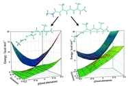
image size:
271.6KB
|
|
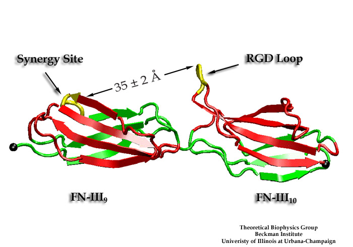
Cells Sense Push and Pull | Mar 2002 |
|
Cells in animals adhere to dynamic, seemingly random assemblies with other
cells that make up tissues like skin, organs, and brain. The cells adhesion
and motion is controlled by the extracellular matrix, with the protein
fibronectin being a key component. The proteins have optimal mechanical
elasticity and also signal to cell surface receptors, integrins, the tension
exerted on them. How this is achieved is the subject of an ongoing
collaboration with the research group of Viola Vogel of the Department of
Bioengineering at the U. of Washington in Seattle (see also Oct 2001
highlight). The most
recent publication
from this effort reports a 97,884 atom steered molecular dynamics simulation using
NAMD. It is revealed now
that stretching two consecutive
domains of fibronectin deforms two sites, the so-called RGD and synergy sites
as well as their distance. This weakens binding to cell receptors and, as a
result, integrins can function as gauges that signal the magnitude of exterior
forces to a cell.
|
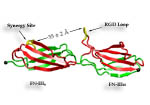
image size:
85.1KB
made with VMD
|
|
Teraflops Harnessed for Biomedical Research | Feb 2002 |
|
Adenosine triphosphate (ATP) is the fuel of life;
every living cell must use ATP to carry out its functions,
and the human body synthesizes its own weight in ATP every day.
The ubiquitous molecular motor ATP synthase catalyzes the
creation of ATP by precisely directing electrochemically generated torque.
A detailed understanding of how this system functions can impact areas
ranging from neurodegenerative disease research to nanotechnology development.
Running at the
Pittsburgh Supercomputing Center
on
LeMieux,
the most powerful computer system in the world for open research,
the freely available simulation code NAMD
can simulate a solvated all-atom model of ATP synthase with full electrostatics
at 65% efficiency on 1000 processors.
This achievement in scalability places NAMD an order of magnitude ahead of
comparable packages for molecular dynamics simulation.
|
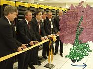
image size:
66.6KB
made with VMD
|
|
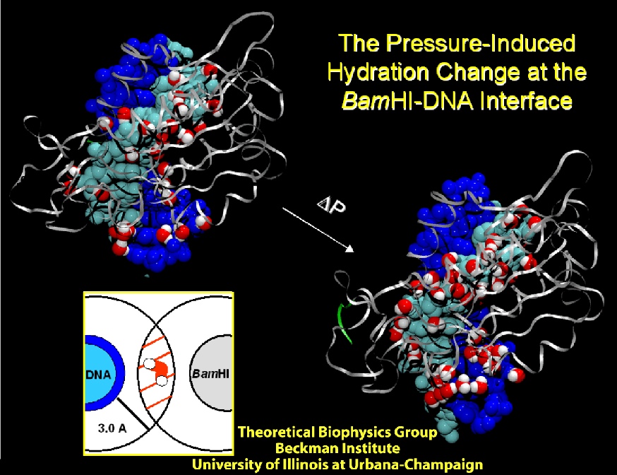
Putting Pressure on Protein-DNA Recognition | Jan 2002 |
|
Deciphering the processes by which proteins recognize and bind to DNA
is critical in our quest to understand cellular functions. To reach
this goal, a collaboration with the group of Stephen Sligar,
UIUC, explored the factors involved in protein-DNA recognition using
hydrostatic pressure to perturb the binding of the BamHI endonuclease
to cognate DNA. Our joint resulting publication
outlines a new technique of high-pressure gel mobility shift analysis
to test the effects of elevated hydrostatic pressure on the binding of
BamHI (so-called restriction enzyme) to a specific DNA sequence. Upon
application of a hydrostatic pressure of 500 bar, recognition between
BamHI and the DNA sequence was weakened nearly 10-fold, suggesting an
important role of water. An advanced 65,000 atom nanosecond molecular
dynamics simulations with NAMD, at both
ambient and elevated pressures, complemented the experiments and
revealed how water-mediated interactions between BamHI and DNA control
sequence recognition.
|
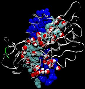
image size:
225.3KB
made with VMD
|
|
|
|
|
| |
|
|
Back to Top
|
Home
This document was last
modified on Thu Jun 27 08:38:02 2002
Material on this page is copyrighted
Contact Webmaster for
more information
6387 accesses since 03 Nov 2000
|
|