
Biologists Confirm Existence of New Cell Structures
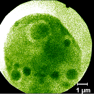
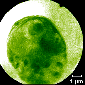
Images of Chlamydomonas, a unicellular green alga, taken with the transmission x-ray microscope on Beamline 6.1. Both images are of the same cell, which was imaged four times. Above is the second of two initial 1-second exposures, showing 1-micron spherical inclusions that are not seen with electron microscopy. Below, the second of two 8-second exposures taken immediately thereafter shows degradation of the inclusions with longer exposure to radiation.
This work was done by A.D. Stead (principal investigator) and T.W. Ford (Royal Holloway, University of London) and W. Meyer-Ilse and J.T. Brown (Center for X-Ray Optics).
Supported by the U.S. Department of Energy under Contract No. DE-AC03-76SF00098.
ALSNews article about this science highlight
More ALS Science